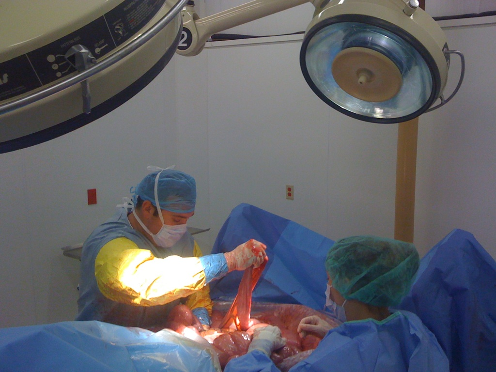
CALEC surgery, or cultivated autologous limbal epithelial cell transplantation, is revolutionizing the landscape of corneal surgery and eye damage treatment. This innovative procedure involves harvesting stem cells from a healthy eye and transplanting them into a damaged eye, effectively restoring the cornea’s surface. A recent clinical trial at Mass Eye and Ear demonstrated that CALEC surgery has a remarkable success rate, offering hope to patients with otherwise untreatable corneal injuries. By utilizing limbal epithelial cells, this groundbreaking approach not only addresses severe vision impairments but also represents a significant advancement in ocular surface repair. As researchers continue to explore the potential of stem cell therapy, CALEC surgery stands out as a beacon of hope for individuals suffering from debilitating eye conditions.
Often referred to as limbal stem cell therapy, CALEC surgery presents an innovative solution for patients facing severe ocular impairments due to corneal damage. This method focuses on the regeneration of limbal epithelial cells, which are crucial for maintaining the eye’s surface integrity. By utilizing harvested stem cells from an unaffected eye, researchers are pioneering a new frontier in eye damage treatment that emphasizes reconstructive techniques and cellular therapy. With promising results in clinical trials, this approach not only enhances visual outcomes but also represents a transformative shift in the methodology of corneal rehabilitation. The quest for effective ocular surface repair continues as the medical community explores the vast potential of this groundbreaking surgical intervention.
Understanding CALEC Surgery: A Breakthrough in Eye Damage Treatment
CALEC surgery, or cultivated autologous limbal epithelial cell therapy, represents a significant advancement in treating severe corneal injuries caused by chemical burns, trauma, or infections. By utilizing healthy stem cells extracted from the patient’s own unaffected eye, this procedure aims to restore the cornea’s surface effectively. The innovation lies in the process, which involves expanding these limbal epithelial cells into a graft that can be transplanted into the damaged eye, thereby revitalizing the ocular surface and potentially restoring vision.
The efficacy of CALEC surgery is underscored by a clinical trial conducted at Mass Eye and Ear, which revealed a remarkable success rate exceeding 90% in restoring corneal surfaces over an 18-month period. This approach not only offers a new hope for individuals with previously untreatable eye damage but also showcases the power of regenerative medicine in ocular health. By harnessing the body’s own stem cells, CALEC surgery provides a personalized treatment option that is both safe and effective, marking a pivotal shift in corneal repair methodologies.
The Role of Stem Cell Therapy in Corneal Surgery
Stem cell therapy has emerged as a revolutionary method in corneal surgery, particularly for patients suffering from debilitating corneal damage. By focusing on the regeneration of limbal epithelial cells—key components in maintaining the cornea’s integrity—this innovative treatment offers a pathway to recovery that traditional methods such as corneal transplants often cannot. The process begins with harvesting healthy stem cells from a donor eye, which are then cultivated in a controlled environment to produce a graft that can be successfully transplanted into the damaged eye.
During the Mass Eye and Ear clinical trial, researchers observed that the stem cell treatment not only restored surfaces but also showed a promising safety profile, with no significant adverse effects reported. The combination of local expertise and advanced biomedical techniques has made it possible to achieve remarkable visual outcomes in a majority of the patients involved. This highlights the potential for further integration of stem cell therapies into mainstream ocular care, opening doors for patients with complex corneal conditions.
The Impact of Limbal Epithelial Cells on Ocular Surface Repair
Limbal epithelial cells play a crucial role in maintaining the health and function of the cornea, acting as a protective barrier that also aids in the regenerative capabilities of the ocular surface. When these cells are compromised due to injury or disease, the integrity of the cornea is threatened, leading to significant vision impairment and discomfort. The restoration of these vital cells through CALEC surgery represents a promising avenue for not only addressing vision issues but also improving the quality of life for affected individuals.
The regeneration of limbal epithelial cells through CALEC surgery does not merely restore the corneal surface; it also facilitates the healing and recovery processes for patients previously left with few treatment options. As ongoing research continues to unveil the mechanisms behind ocular surface repair, the role of CALEC in the wider context of stem cell therapy and eye damage treatment becomes increasingly vital, showcasing the synergy between innovative health solutions and patient-centered care.
Future Prospects for CALEC Surgery: Expanding Treatment Options
The promising initial results of CALEC surgery in treating corneal injuries raise exciting prospects for future advancements in ocular health. The goal of establishing an allogeneic manufacturing process for limbal stem cells sourced from deceased donors could significantly broaden the patient base eligible for such innovative therapies. By making these treatments accessible to individuals with bilateral corneal damage, researchers aim to transform the landscape of eye damage treatment, enabling more patients to regain their vision.
Further studies and clinical trials will be essential to understand the long-term effectiveness and safety of CALEC surgery in various populations. As more data is collected, there is hope for increased interest and investment in this area of research, potentially leading to FDA approval and wider integration in clinical practice. By continuing to refine and expand CALEC applications, the medical community aims to turn this groundbreaking approach into a standard treatment option for those suffering from severe corneal damage.
Innovative Cell Manufacturing Techniques in Ocular Surface Repair
The intricate procedures involved in CALEC surgery are supported by cutting-edge cell manufacturing techniques that have been developed over almost two decades. Ensuring that limbal epithelial cell grafts meet the rigorous quality standards demanded by healthcare authorities is fundamental to the success of the procedure. The collaboration between surgeons and specialized facilities, such as the one at Dana-Farber Cancer Institute, underscores the importance of multidisciplinary approaches in advancing eye damage treatments based on stem cell therapies.
By employing innovative manufacturing techniques, researchers can expand the available quantity of limbal epithelial cells while maintaining their viability and potency for transplantation. The promise of standardized graft production could lead to enhanced treatment availability, which is especially critical for patients with serious ocular surface conditions. This process reflects broader trends in regenerative medicine that emphasize precision, safety, and efficacy in developing treatments for complex health issues.
Evaluating Effectiveness: CALEC Surgery’s Long-term Results
The effectiveness of CALEC surgery is being evaluated through an extensive follow-up framework established during the clinical trials. Participants demonstrated varying degrees of improvement, with a notable percentage achieving complete corneal restoration. These promising outcomes necessitate further assessment to confirm the durability of the effects and establish best practices for long-term management of patients undergoing the procedure.
Understanding the long-term implications of CALEC surgery also involves monitoring possible complications and ensuring that patients receive comprehensive aftercare. This commitment to thorough evaluation not only enhances the treatment process but also contributes valuable data to the scientific community, helping to refine techniques and protocols for future CALEC-related studies.
Safety and Side Effects of Stem Cell-Based Eye Treatments
As with any innovative medical procedure, an assessment of safety and potential side effects is crucial. In the early phases of CALEC surgery trials, a high safety profile was reported, with most adverse events being minor and manageable. This data is reassuring for both patients and healthcare providers, as it highlights the potential of stem cell therapy to transform ocular health without adding significant risk.
Nonetheless, understanding the full spectrum of potential complications is essential as the treatment evolves. Continuous monitoring and reporting on patient experiences will provide invaluable insights into the long-term safety of CALEC surgery, ensuring that any emerging challenges can be effectively addressed and managed.
Regenerative Medicine: The Future of Eye Damage Treatment
Regenerative medicine stands at the forefront of medical advancements, and the application of stem cell therapies like CALEC surgery exemplifies this transformative field. By focusing on the body’s ability to heal and regenerate, these innovations present new solutions for conditions that were once considered hopeless. The successful application of CALEC methodology in corneal surgeries is a prime example of how regenerative therapies can change the prognosis for patients with ocular injuries.
Looking forward, the integration of advanced science with clinical practice holds the potential for developing new treatment modalities that extend beyond corneal issues. As researchers continue to explore the myriad applications of stem cell therapy in various medical fields, the journey of restorative health will likely uncover opportunities for addressing complex injuries and diseases in ways previously unimaginable.
Collaborative Research: A Key to Advancing Ocular Treatments
The collaboration among researchers, surgeons, and various health institutions was instrumental in bringing CALEC surgery from concept to clinical application. This synergy illustrates the necessity of teamwork in advancing medical innovations, particularly in complex fields like ophthalmology. Interdisciplinary partnerships foster the sharing of knowledge and resources, enabling researchers to overcome challenges that may arise during the development of treatments for eye damage.
As CALEC surgery charts its course towards wider adoption, continued collaboration will be vital in addressing the multifaceted aspects of patient care, from initial surgery to long-term follow-up. Such cooperative efforts are likely to enhance the robustness of future studies and pave the way for breakthroughs in other areas of eye health and beyond.
Frequently Asked Questions
What is CALEC surgery and how does it relate to corneal surgery?
CALEC surgery, or Cultivated Autologous Limbal Epithelial Cell surgery, is a pioneering procedure in corneal surgery that uses stem cells to repair and regenerate the corneal surface. This technique involves taking healthy limbal epithelial cells from a donor eye, expanding them into a cellular graft, and transplanting it into a damaged eye. It represents a new hope for treating corneal injuries that were previously irreversible.
How effective is CALEC surgery for treating eye damage?
Recent studies indicate that CALEC surgery has a success rate of over 90% for restoring the corneal surface. In clinical trials, complete restoration of the cornea was achieved in 50% of patients within three months, increasing to approximately 79% and 77% at the 12- and 18-month marks, respectively, demonstrating its effectiveness in treating severe eye damage.
What are the benefits of stem cell therapy in CALEC surgery for ocular surface repair?
Stem cell therapy in CALEC surgery offers significant benefits for ocular surface repair by utilizing the regenerative properties of limbal epithelial cells. This method can restore a healthy corneal surface, alleviate pain, and improve vision in patients suffering from severe corneal injuries caused by trauma, chemical burns, or infections, which are often untreatable by traditional methods.
What types of injuries can CALEC surgery help to treat?
CALEC surgery is designed to treat various types of corneal injuries, particularly those that deplete the limbal epithelial cells, such as chemical burns, infections, or trauma. These injuries can lead to a permanently damaged corneal surface, making CALEC a vital option for restoring eye health and vision in affected patients.
What is the process involved in CALEC surgery for restoring corneal surfaces?
The CALEC surgery process involves several critical steps: first, stem cells are harvested from a healthy eye through a biopsy. These limbal epithelial cells are then expanded into a cellular tissue graft over a period of two to three weeks. Finally, the produced graft is surgically transplanted into the damaged eye, allowing for the restoration of the corneal surface.
Is CALEC surgery currently available for all patients requiring eye damage treatment?
As of now, CALEC surgery remains experimental and is not widely available in the U.S. Further studies are necessary to gather comprehensive data before it can be submitted for federal approval. Currently, it is limited to patients with one affected eye for biopsy purposes, but researchers hope to expand access in the future.
What are potential risks associated with CALEC surgery?
Overall, CALEC surgery has demonstrated a high safety profile with minimal serious adverse events reported. However, potential risks include minor complications like infections, which can occur post-surgery, as seen in one participant attributed to chronic contact lens use. Most adverse events are minor and resolve quickly.
What future developments are anticipated for CALEC surgery and stem cell therapy?
Future developments for CALEC surgery may involve establishing an allogeneic manufacturing process to use limbal stem cells from cadaveric donors. This expansion could enable treatment for patients with damage in both eyes and broaden the application of this promising stem cell therapy for ocular surface repair.
| Key Point | Details |
|---|---|
| First CALEC Surgery | Performed by Ula Jurkunas at Mass Eye and Ear. |
| Purpose of CALEC | To repair corneal damage that was previously untreatable using cultivated autologous limbal epithelial cells. |
| Clinical Trial Results | Showed CALEC was safe and 93% effective for restoring corneal surface after 18 months. |
| Procedure Overview | Involves harvesting stem cells from a healthy eye, expanding them into grafts, and transplanting into the damaged eye. |
| Future Implications | Plans to establish an allogeneic process for broader treatment possibilities. |
| Research and Funding | Funded by the National Eye Institute, the trial is the first human study of stem cell therapy for the eye in the U.S. |
Summary
CALEC surgery introduces a groundbreaking approach to treating corneal injuries that were once considered irreparable. Ula Jurkunas and her team have made incredible strides in utilizing stem cells from healthy eyes, demonstrating impressive recovery rates in patients with corneal damage. While CALEC surgery is still in the experimental phase, its high efficacy and safety profile present a promising future for individuals suffering from irreversible corneal defects. Ongoing research and advancements could soon make CALEC a standard treatment, allowing broader access to vital ocular restoration therapies.


